powered by
Ankle & Foot Ultrasound
- SAME Day or NEXT Day Appointments
- FREE consultation to make sure this is the right scan for you
- Performed by a CONSULTANT RADIOLOGIST
- PRICE: £175

An ultrasound scan of the ankle & foot is performed to have a look at the ankle joint and muscles surrounding the ankle. It is also possible to see some of the ligaments, in particular the Achilles tendon, calcaneo-fibular ligament (CFL) and the anterior talo-fibular ligament (ATFL).
Common conditions
An ankle & foot ultrasound scan is useful in evaluating the following conditions:
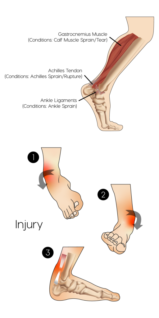
The achilles tendon attaches the gastrocnemius muscle (main calf muscle) to the calcaneus (heel) bone. The gastrocnemius muscle helps with jumping and pushing the foot down.
Following overuse, especially in sports such as badminton, the tendon can become inflamed and sore. At this point this is referred to as tendinitis. Trying to tip toe and even walk can become quite difficult and even painful. An ultrasound scan can help to diagnose an achilles tendinitis and also differentiate it from an achilles tendon rupture.
The achilles tendon attaches the gastrocnemius muscle (main calf muscle) to the calcaneum (heel) bone. The gastrocnemius muscle helps with jumping and pushing the foot down.
The achilles tendon can suffer a rupture (tear) during intense periods of sport or exertional activity. It often occurs suddenly and can feel like someone has ‘kicked you’ at the back of your heel. This will occur as you are about to push off on your feet to either run or jump and is a common injury in sports such as football and badminton. It is normally not possible to walk after such an injury.
An ultrasound scan can diagnose and confirm the presence of an achilles tendon rupture. It can also help to differentiate between a partial and complete rupture – this information has significant consequences regarding treatment and length of recovery.
The treatment of an achilles tendon rupture consists of placing the foot, ankle and lower leg in a special plaster cast called an equinus cast. Often patients are left in this cast for a
period of 8 to 10 weeks with regular adjustments to the foot and ankle portion. Surgical repair is also an option and recommended for those who are especially active and play a lot of sports. The recovery period for a surgical repair is just as long and also still requires an equinus cast. Those receiving a surgical repair have a lower rate of future re-rupture as opposed to those who are just treated with a plaster cast.
An ankle sprain is an extremely common injury. The most common mechanism of injury is an inversion injury. This is when the foot bends inwards. Eversion injuries are less common and occur when the foot bends outwards.
Ankle sprains often heal up by themselves following a period of rest and limited weight bearing on the ankle. Crutches may be needed. However, in some cases, an ankle sprain can take a long time to improve, in which case it can help to perform an ultrasound scan to take a look at the ligaments that hold the ankle in place and give it stability.
The ATFL is also known as the Anterior TaloFibular Ligament and runs from the ankle to the forefoot. This can often be injured in an inversion injury. The ATFL can be seen on ultrasound and any tears identified.
The CFL is also known as the CalcaneoFibular Ligament and runs from the calcaneus (heel bone) to the back and side of the ankle. This also can be injured during an inversion injury. Ultrasound can be used to identify any significant injuries to the CFL following an ankle sprain.
The calf consists of two main muscles. The bulk of the calf comprises the gastrocnemius muscle. This lies above the deeper soleus muscle. The gastrocnemius muscle has two bellies which attach to either side of the knee joint above and via the achilles tendon to the heel below. The gastrocnemius helps with running and jumping and is the dominant muscle allowing you to push off with your feet.
All high impact sports that consist of running and jumping are heavily reliant on the gastrocnemius muscle. Overuse, sudden physical activity without warm ups and warm downs as well as direct trauma can result in minor and major tears to the gastrocnemius. This can make it difficult to walk let alone run. Ultrasound is the first port of call in diagnosing these injuries and can help provide a timeline for recovery. Healing of such injuries can be accelerated through the use of Magellan plasma therapy administered under ultrasound guidance. Both of these services are available at Rejuvence Scans.
Risks
Ultrasound scan of the ankle & foot is a safe procedure and has no known risks.
How to prepare
No specific preparation is required for this scan.
What you can expect
Before your ultrasound, you may be asked to change into a gown and to remove any jewelry. You will be asked to sit on an examination table.
A radiologist will perform your scan. A small amount of ultrasound gel is applied to your ankle. The gel enables the ultrasound device to provide better images.
The radiologist will gently press an ultrasound probe against various points on your ankle, lower leg and foot. They will also ask you to move your foot into different positions. Depending upon your symptoms you may experience some pain during this scan. The radiologist will always try to make the scan as comfortable as possible. If you take regular pain medication please have these to hand when you have your scan as you maybe a little sore afterwards. The ultrasound scan takes around 30 minutes to complete.
You will be able to return to normal activities immediately after your scan.
Results
The radiologist will prepare a written report immediately after your scan. You can wait for the written report and should you wish a copy of your scan images can be sent to you via email so you have them to hand at all times.
Ultrasound guided joint & muscle injections
Following identification of certain injuries or conditions we are also able to offer ultrasound guided cortisone (steroid) injections. Cortisone injections help to reduce inflammation and if combined with local anaesthetic can provide significant pain relief for upto 6 weeks.
Using the Magellan system ultrasound guided biocellular injections can help to accelerate the repair of muscle injuries. This is a treatment elite athletes often use to come back from injury quicker.
Follow up with Rejuvence Scans
We always recommend booking in a consultation immediately after your scan with one of our doctors to discuss the results of your scan and to provide advice regarding any further investigations and/or treatment. Further investigations and treatment can include:
Blood tests
Referral to a specialist (Private/NHS*)
Referral for further imaging (Private MRI)
* Please note that for referrals back to NHS you will still have to go via your GP but Rejuvence Medical will provide a full report and cover letter in support of the referral.