powered by
Deep Vein Thrombosis
- SAME Day or NEXT Day Appointments
- FREE consultation to make sure this is the right scan for you
- Scan of BOTH legs included.
- Performed by a CONSULTANT RADIOLOGIST
- COST: £165.00
At Rejuvence Scans we offer a comprehensive, efficient and professional medical ultrasound scanning service. We are registered with the Care Quality Commission (CQC). Our radiologists have years of NHS experience and we have a robust vetting and checking service provided by a network of radiologists. We can also arrange for you to take away a copy of your ultrasound scan.
A deep vein thrombosis scan is performed to search for evidence of a clot in the veins of the legs and thighs.
Risks
Symptoms


Conditions
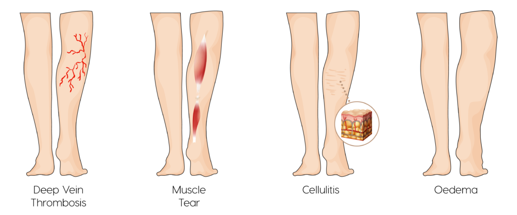
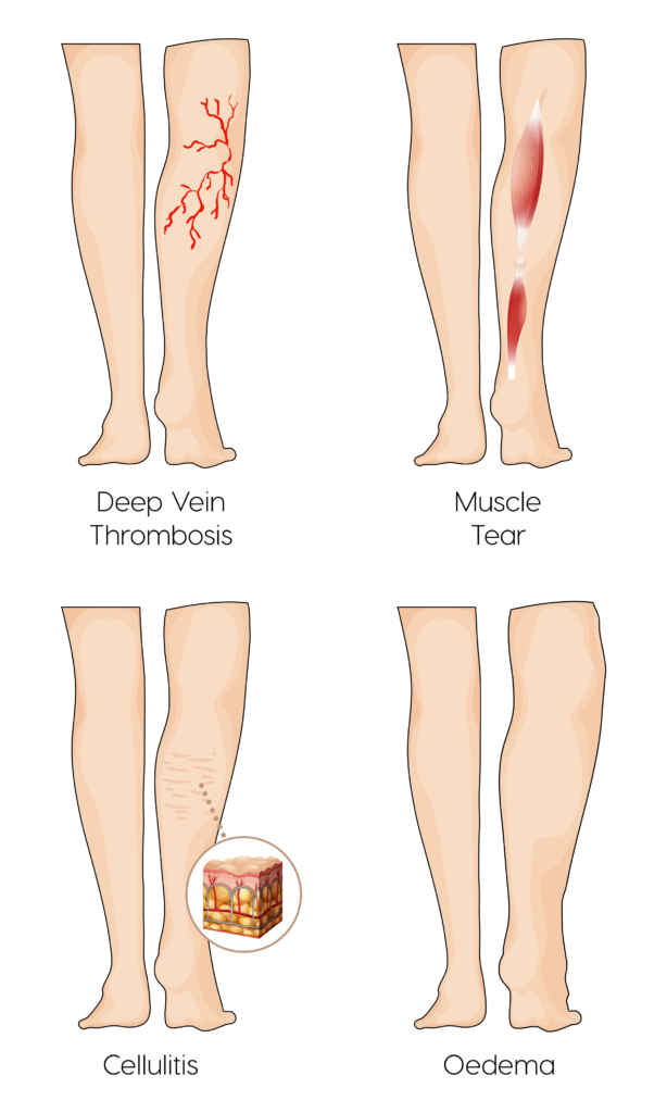
The word thrombosis means blood clot. The legs and thighs consist of a complex network of veins that carry blood back up to the heart. This network of veins can be broadly categorized into superficial veins and deep veins. A deep vein thrombosis is a blood clot that develops one of the deep veins of the legs.
The probability of developing DVT is approximately 1 in 1000 per year. However, certain factors greatly increase this risk. Young people have less of a chance of developing a DVT than the elderly. The cumulative chance of developing DVT over a lifetime ranges from 2 percent to 5 percent. There are an estimated 300,000 first-time DVTs diagnosed in the US.
Men appear to have a greater risk of developing DVTs than women but women are susceptible to more risk factors.
All ages can be affected by DVTs although the risk in those aged less than 18 years appears to be extremely low and this risk increases as we age and is significantly higher in the elderly.
All ages can be affected by DVTs although the risk in those aged less than 18 years appears to be extremely low and this risk increases as we age and is significantly higher in the elderly.
A deep vein thrombosis is often the result of reduced flow of blood through the deep veins. Blood has a natural tendency to clot and hence if the flow of blood is sufficiently reduced it will form clots that can cause an obstruction and hence a DVT. There are a number of risk factors that can predispose someone to developing a DVT:
- Prolonged in-activity: Significantly reduced levels of activity and movement can increase the risk of DVT. Examples include during a long haul flight, bed rest after surgery and reduced activity generally due to paralysis, obesity or other disabilities. Certain occupations such as long haul driving or sitting can also increase DVT risk.
- Obesity: Being overweight on it’s own can increase the risk of developing a DVT due to increased pressure on the veins of the pelvis and legs.
- Oral Contraceptive Pill and Hormone Replacement: Regular use of birth control pills and hormone replacement therapy especially after the menopause or associated with treatments following breast or ovarian cancer are associated with a slightly increased risk of developing DVTs.
- Surgery: Long surgical procedures, especially abdominal and pelvic surgery, are associated with significantly increased risks of DVTs. For this reason many of these procedures require patients to take preventative doses of blood thinning medication to mitigate this increased risk.
- Pregnancy: There are significant physiological changes during pregnancy that often result in increased pressure on the veins of the pelvis and legs. This results in an increase of developing a DVT. This risk continues into the postpartum period as well for 6 weeks after giving birth.
- Inherited Clotting Disorders: The presence of inherited clotting disorders that result in a greater tendency for blood to form clots increases the risk of developing DVTs. Such conditions include Protein S and Protein C deficiency.
- Family history of DVT: The presence of family members who have suffered from DVTs can increase your own risk of developing a DVT. Although on its own, normally not a significant risk, when combined with any of the above risks, the rate of DVT is much higher.
- Cancer: The presence of cancer significantly increases the risk of developing a DVT.
The most common presentation of a DVT is in the lower limbs although it is theoretically possible to develop a DVT in the upper limbs as well. Most develop sudden onset swelling to the lower leg. This can be accompanied by worsening pain in the calf and thigh depending on the position of the DVT within the deep venous system. Associated redness and infection can arise subsequently in the form of what is known as thrombophlebitis. Where a large DVT has developed the legs may be very swollen and there is an increased risk of the clot breaking off and lodging in the lungs causing a pulmonary embolism. If one develops chest pain, shortness of breath or starts coughing up blood alongside swelling in the leg, it is especially important to seek urgent medical attention. You should immediately arrange to see a doctor.
Ultrasound is the investigation of choice for identifying a DVT especially in the lower legs and thighs. It is a quick and very accurate scan. Prior to getting an ultrasound scan certain blood tests can also be performed in association with specific risk stratification scores to work out whether an ultrasound scan is needed. This is because many other conditions can also present in a similar fashion to DVTs. Hence, where risk factors are not clearly present, blood tests such as a D-Dimer can be performed alongside a Well’s Score to ascertain whether blood thinning treatment should be started and if an ultrasound scan should subsequently be arranged.
For those at high risk of developing a DVT, preventative lifestyle measures are advised including minimising periods of inactivity and wearing tight compression stockings to aid with movement of blood through the deep veins of the legs. In high risk cases it may be necessary to take preventative doses of blood thinning medication for a specific period of time.
A deep vein thrombosis is normally broken down and removed by the body itself. The importance of treatment in most cases is to do with stopping the clot from getting larger and breaking off to cause blockages in blood flow further down, particularly in the lungs where it can cause a pulmonary embolism.
The majority of patients are immediately started on blood thinning medication in the form of heparin, warfarin or a NOAC. This medication is normally recommended for use for a minimum period of 6 months. In certain cases a longer period of treatment is required and this could even be lifelong. Where the DVT is particularly large and lodged in deeper and larger veins such as the common iliac vein, specific procedures may be recommended to break up the clot with clot busting medication or even have it removed through a surgical procedure – embolectomy.
Risks
Deep Vein Thrombosis (DVT) ultrasound is a safe procedure and has no known risks.
How to prepare
No specific preparation is needed for this scan.
Please keep in mind that DVT is a potentially serious condition. If you have not been seen by a medical professional and not been started on blood thinning medication prior to your scan we would recommend you book an urgent appointment with one of our doctors to assess your symptoms and make a judgement on how likely it is that you have a DVT. Treatment should be started without delay regardless of whether the diagnosis has been confirmed or not. Blood thinning medication can easily be stopped if it turns out you do not have a clot.
What you can expect
Before your ultrasound, you may be asked to change into a gown and to remove any jewelry. You will be asked to lie on your back.
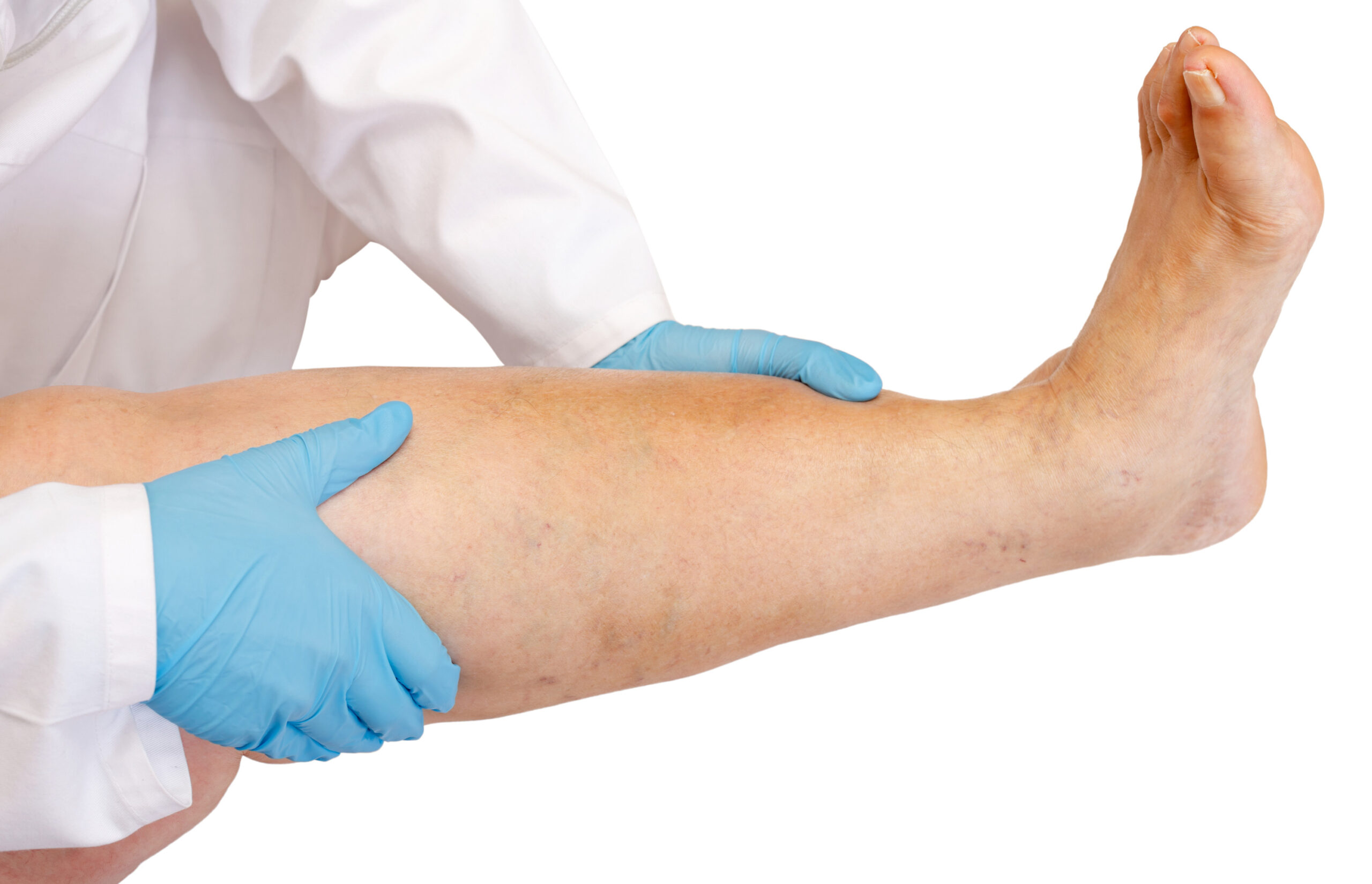
A radiologist will perform your scan. A small amount of ultrasound gel is applied to your thigh and lower leg. The gel enables the ultrasound device to provide better images.
The radiologist will gently press an ultrasound probe against your lower limb, moving it back and forth along the veins. A standard ultrasound scan takes around 20 minutes to complete. It is not uncommon to experience some discomfort during this scan as the radiologist is required to press firmly on your deep veins to check if they are compressible. Lack of compression can suggest the presence of a deep vein thrombosis.
You will be able to return to normal activities immediately after your scan.
Results
The radiologist will prepare a written report immediately after your scan. Should your scan confirm the presence of DVT you should be immediately started on blood thinning medication if this has not already been done.
If the scan is negative blood thinning medication can be stopped.
Both these steps should be carried out in conjunction with your own GP or one of our doctors at Rejuvence Scans. At Rejuvence Scans we will always make a doctor available after a DVT scan.
Follow up with Rejuvence Scans
We always recommend booking in a consultation immediately after your scan with one of our doctors to discuss the results of your scan and to provide advice regarding any further investigations and/or treatment. Further investigations and treatment can include:
Blood tests
Referral to a specialist (Private/NHS*)
Referral for further imaging (Private MRI)
* Please note that for referrals back to NHS you will still have to go via your GP but Rejuvence Medical will provide a full report and cover letter in support of the referral.