powered by
Kidney Ureter Bladder (KUB) Ultrasound
- SAME Day or NEXT Day Appointments
- FREE consultation to make sure this is the right scan for you
- Performed by a CONSULTANT RADIOLOGIST
- COST: £165.00
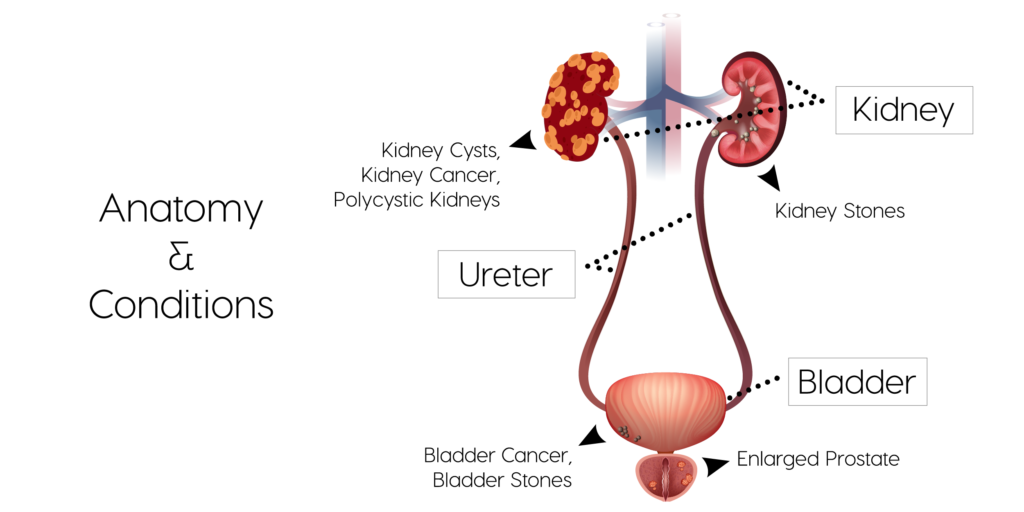
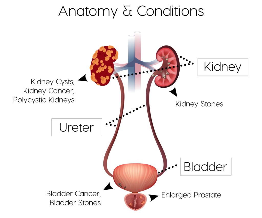
A Kidney Ureter Bladder (KUB) ultrasound is performed to have a look at the kidney, ureter (tubes connecting the kidney to the bladder) and the bladder.
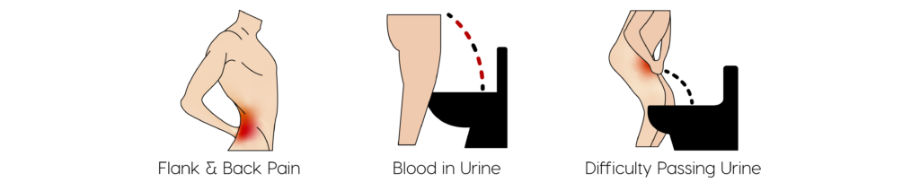
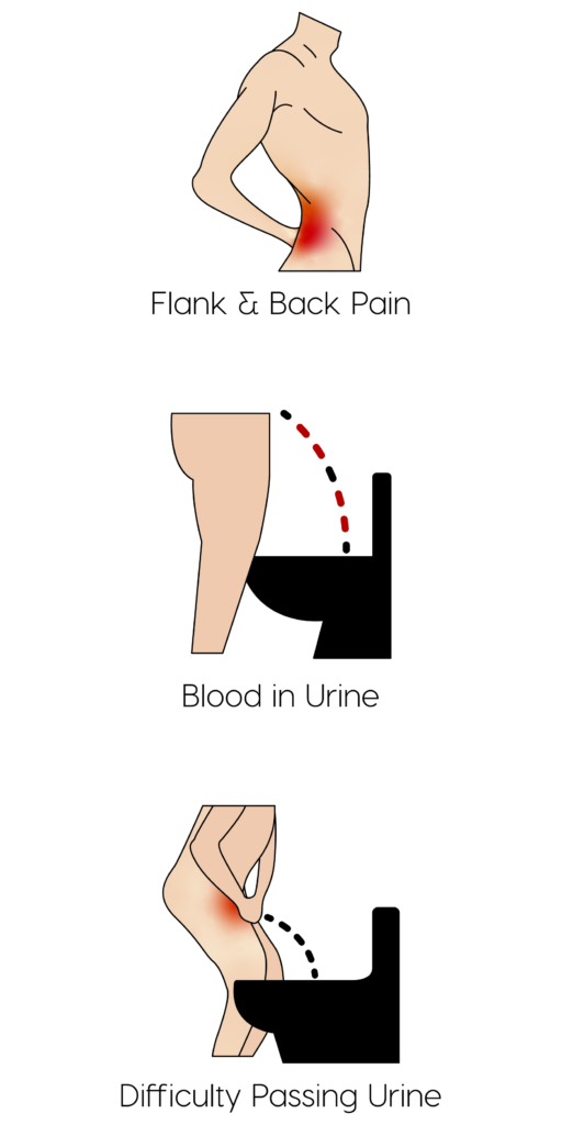
Symptoms
Common conditions
A KUB ultrasound scan is useful in evaluating the following conditions and structural changes in the kidney, ureter and bladder:
- What is Hydronephrosis? Hydronephrosis describes abnormal swelling of the kidney. This is usually the result of obstruction of the ureter (tube draining urine from the kidney).
- How common is it?
Although this is most common in babies, it can also affect adults, and, especially pregnant women. - Does it affect males and females?
Is more common in women, but it also appears in men with prostatic hypertrophy and prostate cancer. - What age groups?
Patients between the ages of 20-60 years old are commonly affected. - What are the main symptoms?
Most commonly, patients will experience pain on either side of the back, abdomen or groin, pain while urinating, an increase in the need to urinate, incontinence or the inability to completely empty the bladder whilst passing urine. - How is it tested for?
Blood tests can be used to provide an indication of how well the kidneys are working. Urine tests help to identify the presence of any urine infections. Ultrasound is diagnostic for identifying hydronephrosis. - Is there a cure?
It is a curable condition that can be resolved through various treatment options, but in the most severe cases it could lead to kidney failure and permanent damage. - How is it treated?
Treatment is very much dependent upon the cause of the swelling of the kidney. Kidney stones blocking the ureter can be removed either using lithotripsy or surgically. Infections can be treated with antibiotics. In severe cases, especially when kidney function is compromised it may be necessary to insert a nephrostomy tube to directly drain the kidney.
- What is hydroureter?
Hydroureter is swelling of the ureter that can be normal or abnormal. It varies in severity from acute, chronic, on one side or both sides. It can also be the result of a blockage somewhere in the urinary system.
- How common is it?
When there is a urinary tract infection or condition affecting the kidneys or bladder, ureteral obstruction can be common and due to its treatability, there are rarely any severe complications associated with hydroureter.
- Does it affect males and females?
Hydroureter occurs more commonly in women, a large percentage of them being pregnant.
- What age groups?
Most often patients between the ages of 20-60 will be affected. In men, prostatic diseases cause a higher risk for developing hydroureter after the age of 60.
- What are the main symptoms?
Signs of hydroureter and ureteral obstructions may include pain in the flanks of the back, lower abdomen or groin, a change in the amount of urine that is produced, having a hard time urinating or having painful urination. It is also common to see blood in the urine and have high blood pressure. Those who have had previous urinary tract infections are at higher risk for developing hydroureter.
- How is it tested for?
A blood test can be done to assess the functioning of the kidneys as well as a urine test to check for any signs of a possible infection or blood suggesting the presence of kidney stones that may be causing a blockage. Another common method for diagnosis of hydroureter is through ultrasound.
- Is there a cure?
There are many treatments available for hydroureter that can be administered as long as the condition is assessed early and before it has the chance to cause any major damage to the kidneys or urinary tract.
- How is it treated?
Hydroureter is often the result of various possible conditions affecting the kidneys. Treatment is dependent upon the underlying cause.
- What is a renal cyst?
Kidney cysts are fluid filled sac like structures found within the kidney itself.
- How common is it?
Kidney cysts are relatively common.
- Does it affect males and females?
Kidney cysts are common in both males and females.
- What age groups?
Kidney cysts can occur at any age but are often identified more commonly in those aged 40 years and older.
- What are the main symptoms?
On the whole a single kidney cyst does not cause any symptoms unless it is extremely large in which case it can cause fullness and an ache in the corresponding flank. If the cyst ruptures there can be severe pain and associated blood in the urine.
- How is it tested for?
A kidney cyst can easily be identified following an ultrasound scan. They can also be seen on CT scans and MRI scans. They are normally incidental findings reported whilst performing a scan for a different reason.
- Is there a cure?
There is no specific cure for kidney cysts.
- How is it treated?
Single or a couple of kidney cysts normally do not require any specific treatment. They normally do not affect kidney function. In situations where the cyst needs to removed surgery is required.
- What is Polycystic Kidney Disease?
PKD (polycystic kidney disease) is a kidney disorder than is often inherited. It causes multiple cysts filled with fluid, to develop in the kidney and can cause problems in the functionality of the kidneys, possibly leading to eventual kidney failure.
- How common is it?
As far as genetic disorders go, PKD is one of the most common and affects around 500,000 people in the USA.
- Does it affect males and females?
The disease tends to occur more in women than men, affecting more Caucasians than African Americans.
- What age groups?
Most patients will often develop the disease between the ages of 30-40, but it can show signs of development in children as well.
- What are the main symptoms?
Commonly patients will experience high blood pressure, headaches and back pain. They may also feel a fullness or swelling in the abdomen due to the enlarged kidneys. The passing of kidney stones can also be a sign of PKD, as well as seeing blood in the urine, frequent urine or kidney infections and, in serious cases, kidney failure.
- How is it tested for?
Polycystic Kidney Disease is easily diagnosed on abdominal ultrasound.
- Is there a cure?
There is no known cure for PKD; however, there are treatments that are used to reduce damage to the kidneys.
- How is it treated?
In severe cases of kidney dysfunction kidney transplant maybe needed.
- What are Kidney Stones?
Kidney stones are hard little “pebbles” that can form when a patient’s urine contains crystal-forming salts or minerals like calcium, oxalate or uric acid. If the urine is unable to dilute these salts or crystals, they can stick together to form kidney stones that can be as small as a grain of sand or even as large as a golf ball.
- How common is it?
Generally speaking, most people don’t have to worry about kidney stones too much. They can be more common in people with high blood pressure, diabetes and obesity.
- Does it affect males and females?
Kidney stones are more common in men by about 10%. 19% of men and 9% of women find themselves affected by the stones.
- What age groups?
Kidney stones can develop at any time in a person’s life, sometimes even in premature babies. Kidney stones are most common in men between the ages of 20-50.
- What are the main symptoms?
A person might not know they have a kidney stone until is moves in the kidney or begins to pass into the ureter. When this happens, the most common symptoms include severe pain occurring in the sides and back just below the ribs. This pain can come in waves and be moderate to extreme. Whilst the stone is trying to pass it will likely cause pain during urinating and the color of the urine may be pink, red or brown. Alongside severe pain they can also be associated with nausea and vomiting. Kidney stones are associated with an increased risk of kidney infections such as pyelonephritis.
- How is it tested for?
CT scanning if the gold standard for diagnosing and identifying kidney stones. Ultrasound in skilled hands can be used to identify kidney stones. Ultrasound can also identify hydroureter developing as a result of an obstructing kidney stone.
- Is there a cure?
Kidney stones, depending on their size, often pass on their own through the urinary tract. Where larger stones have been identified these will need addressing. Changes to diet and medication can help to reduce the rate of kidney stone formation.
- How is it treated?
Where kidney stones are relatively large and unlikely to pass on their own or indeed causing an obstruction to the urinary tract there are specific treatment options. A stent can be passed into the ureters to help drain kidneys that are obstructed. Lithotripsy uses sound waves to break up kidney stones. In very large stones surgery maybe necessary.
- What is Kidney Cancer?
There are many forms of kidney cancer, renal cell carcinoma being the most common. When healthy cells in the kidney or both kidneys begin to grow out of control, they can form a tumor. The renal tubules clean your blood and produce urine. Generally, renal cell cancer will remain in the kidney or kidneys, but it has been known to spread to the bones, lungs and brain.
- How common is it?
In 2019 the American Cancer Society estimated that out of 73,820 new cases of kidney cancer, sadly, about 14,770 of them will be fatal. The rate of new kidney cancer cases has been growing since the 90’s.
- Does it affect males and females?
Kidney cancer is the sixth most common cancer that occurs in men and the eighth most common that occurs in women. Men are more likely to develop kidney cancer in their lives though the disease is common in both men and women.
- What age groups?
On average, kidney cancer is generally uncommon in patients younger than 45 and occurs most commonly in patients in their mid-sixties.
- What are the main symptoms?
The most common symptom of kidney cancer is seeing blood in the urine. There is also likely to be a pain in the back or flank of the abdomen. More rarely a mass can be felt in the flank. Other symptoms include swelling of the legs and ankles, high blood pressure, low red blood cell count (known as anemia), fatigue, night sweats, weight loss, loss of appetite, fever and men can often experience a swollen vein in the scrotum.
- How is it tested for?
An initial ultrasound scan of the kidney can identify an abnormal mass in the kidney. This can be further defined using CT or MRI. Blood tests are also used to further confirm the diagnosis followed by biopsy under CT guidance.
- Is there a cure?
The only way to definitively cure cancer is to remove it entirely. If it can be detected early then yes, kidney cancer can be cured in most cases. The overall chances of a full recovery from kidney cancer are good.
- How is it treated?
First line treatment is a nephrectomy – complete removal of the affected kidney. Depending upon whether the kidney cancer has spread and associate with metastases, a combination of chemotherapy and radiotherapy may be necessary.
- What is Bladder Cancer?
Bladder cancer is the growth of a tumor in the bladder lining which can, in some cases, spread to the muscles of the bladder. It is characteristically associated with azo dye use in the leather tanning industry.
- How common is it?
As one of the most common cancers, bladder cancer occurs in approximately 68,000 adults in the US yearly.
- Does it affect males and females?
Though bladder cancer affects both men and women, it occurs most often in men.
- What age groups?
Anyone can develop bladder cancer at any age; however, it tends to occur more often in older adults in their late 60’s.
- What are the main symptoms?
The most common sign of bladder cancer is painless haematuria (blood in the urine). It can also be associated with incomplete emptying of the bladder and lower urinary tract infections.
- How is it tested for?
Doctors will commonly perform a cystoscopy in which a small tube is inserted into the bladder through the urethra. The bladder line is inspected and biopsies taken. Ultrasound can also help to identify bladder cancer.
- Is there a cure?
Bladder cancer can be completely cured through early detection and treatment.
- How is it treated?
Surgery can be done to remove the tumors if the conditions are favorable, however, if the bladder must be removed completely then reconstructive surgery is needed to be able to pass urine. Follow up with different forms of chemotherapy and radiotherapy may also be needed.
- What is an Enlarged Prostate and Prostate Cancer?
The prostate is a gland found in men located just below the bladder.
Enlarged Prostate – simple enlargement of the prostate gland. - Prostate Cancer – following excessive enlargement of the prostate it can become cancerous.
- How common is it?
Prostate cancer is one of the highest occurring cancers in men. An enlarged prostate is also very common in men over the age of 50. - What age groups?
An enlarged prostate generally occurs in men between 51 and 60 years. It occurs in upto 90% of men above the age of 80. Prostate cancer most commonly affects men over the age of 50 and of Afro-Carribean descent. - What are the main symptoms?
In an Enlarged Prostate men can experience weak/slow urine flow, feelings of incomplete urination and difficulty in starting urination. There is also increased frequency and urgency of urination (needing to go more often with difficulty holding urine). The same symptoms are experienced with prostate cancer as well as blood in the urine, blood in the semen, and the onset of erectile dysfunction. - How is it tested for?
A doctor may perform a digital rectal exam to check for prostatic enlargement. Urine tests can also help. Blood tests such as a prostate-specific antigen (PSA) blood test is usually the first line of investigation followed by biopsy. - Is there a cure?
There are several treatment options for an enlarged prostate. For prostate cancer early detection is imperative for a good outcome. - How is it treated?
Medication such as alpha-blockers to relax the prostate and bladder muscles can be used to help with passing urine. A TURP (trans-urethral resection of the prostate) can be performed to shave away part of the prostate and reduce it in size. For prostate cancer a TURP maybe sufficient. However, in more advanced cases the entire prostate may need to be removed (prostatectomy) and this is coupled with a combination of chemotherapy and radiotherapy.
Risks
KUB ultrasound is a safe procedure and has no known risks.
How to prepare
Ideally you should avoid food for 8 hours prior to your scan. We understand that this may of course not be practical especially if you are coming from work. Therefore, if possible please try to skip lunch. Food in your stomach can make it difficult for the radiologist to generate clear images. The radiologist will also request that you try to have a full bladder prior to your scan. Drink plenty and avoid going to the toilet immediately prior to your scan. In general you should continue to take your regular medication.
What you can expect
Before your ultrasound, you may be asked to change into gown and to remove any jewelry. You will be asked to lie on your back.
A radiologist will perform your scan. A small amount of ultrasound gel is applied to your abdomen. The gel enables the ultrasound device to provide better images.
The radiologist will gently press an ultrasound probe against your tummy, moving it back and forth. A standard ultrasound scan takes around 20 minutes to complete. It’s usually painless. However, it is not uncommon to experience some discomfort if radiologist is required to press down on areas where you may have some pain. We always try to ensure you are comfortable at all times.
The radiologist will gently press an ultrasound probe against various points on your knee. They will also ask you to move your leg into different positions. Depending upon your symptoms you may experience some pain during this scan. The radiologist will always try to make the scan as comfortable as possible. If you take regular pain medication please have these to hand when you have your scan as you maybe a little sore afterwards. The ultrasound scan takes around 30 minutes to complete.
You will be able to return to normal activities immediately after your scan.
Results
The radiologist will prepare a written report immediately after your scan. You can wait for the written report and should you wish a copy of your scan images can be sent to you via email so you have them to hand at all times.
Follow up with Rejuvence Scans
We always recommend booking a consultation immediately after your scan with one of our doctors to discuss the results of your scan and to provide advice regarding any further investigations and/or treatment. Further investigations and treatment can include:
Blood tests
Urine dipstick
Referral to a specialist (Private/NHS*)
Referral for further imaging (CT/MRI)
* Please note that for referrals back to NHS you will still have to go via your GP but Rejuvence Medical will provide a full report and cover letter in support of the referral.