powered by
Knee Ultrasound
- SAME Day or NEXT Day Appointments
- FREE consultation to make sure this is the right scan for you
- Performed by a CONSULTANT RADIOLOGIST
- PRICE: £175
Symptoms

An ultrasound scan of the knee is performed to have a look at the knee joint and muscles surrounding the knee. It is also possible to see some of the ligaments and the mensici within the knee joint. The quadriceps and patella tendon are also examined during this scan.
Anatomy & conditions
Common conditions
A knee ultrasound scan is useful in evaluating the following conditions:
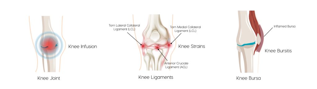
- What is Knee Effusion?
Knee Effusion is most commonly known as fluid on the knee and happens when excess fluid accumulates in or around the knee joint.
- How common is it?
It is a relatively common condition.
- Does it affect males and females?
Both men and women can be affected by knee effusion.
- What age groups?
Knee effusion can occur in people of any age but is most common.
- What are the main symptoms?
The main symptoms of knee effusion are swelling and redness of the skin around the kneecap, stiffness in the joint and difficulty straightening or putting weight on the knee, alongside a painful knee.
- How is it tested for?
Testing for knee effusion can be done through a physical exam where the doctor may apply pressure with their hands to feel for the presence of fluid on the kneecap. - How is it treated?
It is treated with rest, ice, compression and elevation. A patient can also take anti-inflammatory medications to reduce pain and swelling.
- What is a Knee Sprain?
A knee sprain results when knee ligaments are torn or overstretched causing pain and restriction of movement. - How common is it?
Knee sprains are common during sports that involve a lot of running, jumping and turning. - Does it affect males and females?
Knee sprains can affect both men and women. - What age groups?
People between the ages of 15-24 years old are the most likely to suffer from a higher rate of sprained knees. However the majority of knee sprains happen in people aged 25-44 years old. - What are the main symptoms?
A knee sprain may cause symptoms such as stiffness or decreased movement, pain and tenderness, a popping that you can hear or feel in the knee, swelling and bruising and a knee that will buckle or give out when trying to walk. - How is it tested for?
A doctor can perform a physical exam to determine if a knee is sprained by evaluating pain and clicking or popping in the joint. - How is it treated?
Minor knee sprains are likely to heal over time with rest, ice, compression and elevation. Some people may also take anti-inflammatories and aspirin for relief of pain and swelling. Significant ligament tears will require surgery.
- What is Patella Bursitis?
Patella Bursitis is an inflammation of the bursa in the front, above or below the kneecap which takes place when too much fluid causes it to swell and adds pressure to parts of the knee. - How common is it?
Patella Bursitis can be common in people with repetitive work tasks that result in resting on the knees. - Does it affect males and females?
It can affect both men and women though it tends to occur more commonly in men. - What age groups?
The most common age group affected by patella bursitis are men between the ages of 40-60 years. - What are the main symptoms?
Common symptoms of patella bursitis are pain during activity, rapid swelling in the front of the kneecap, tenderness and a slight fever to the area. In more severe cases where an infection is present a fever and chills may be experienced. - How is it tested for?
Testing is commonly done through a physical exam of the patient’s knee joint looking for swelling and pain as well as range of motion. Aspiration and lab tests may also be required. - How is it treated?
Doctors will suggest rest, ice, compression and elevation of the knee as well as anti-inflammatory medications to reduce pain and swelling. It can also be helpful to remove the bursa fluid.
- Housemaid’s Knees is also known as pre-patellar bursitis. Housemaid’s Knees occurs following excessive time spent resting on your knees during activities such as scrubbing a floor. Certain occupations are particularly prone to Housemaid’s Knees such as plumbers and electricians. Symptoms include pain and swelling directly over the knee cap and is the result of prepatellar bursitis. It can be identified through an ultrasound scan. Treatments include anti-inflammatory painkillers and in severe cases excessive fluid build up may need to be drained. Prevention is better than cure hence the use of knee pads is recommended.
Clergy Man’s Knees is also known as infra-patellar bursitis. It typically occurs following excessive pressure on the suprapatellar bursa due to long periods of time in the classic prayer position. Symptoms include pain and swelling just below the knee cap. Treatments include anti-inflammatory painkillers and in severe cases excessive fluid build up may need to be drained.
Risks
Ultrasound scan of the knee is a safe procedure and has no known risks.
How to prepare
No specific preparation is required for this scan.
What you can expect
Before your ultrasound, you may be asked to change into a gown and to remove any jewelry. You will be asked to sit on an examination table.
A radiologist will perform your scan. A small amount of ultrasound gel is applied to your knee. The gel enables the ultrasound device to provide better images.
The radiologist will gently press an ultrasound probe against various points on your knee. They will also ask you to move your leg into different positions. Depending upon your symptoms you may experience some pain during this scan. The radiologist will always try to make the scan as comfortable as possible. If you take regular pain medication please have these to hand when you have your scan as you maybe a little sore afterwards. The ultrasound scan takes around 30 minutes to complete.
You will be able to return to normal activities immediately after your scan.
Results
The radiologist will prepare a written report immediately after your scan. You can wait for the written report and should you wish a copy of your scan images can be sent to you via email so you have them to hand at all times.
Follow up with Rejuvence Scans
We always recommend booking in a consultation immediately after your scan with one of our doctors to discuss the results of your scan and to provide advice regarding any further investigations and/or treatment. Further investigations and treatment can include:
Blood tests
Referral to a specialist (Private/NHS*)
Referral for further imaging (Private MRI)
* Please note that for referrals back to NHS you will still have to go via your GP but Rejuvence Scans will provide a full report and cover letter in support of the referral.
Ultrasound guided drainage of bursitis
For immediate symptomatic relief it is possible to perform ultrasound guided drainage of large swellings around the elbow. The fluid drained can also be sent off to the lab to identify the presence of any infection. Following drainage a course of antibiotics will normally be prescribed.
Ultrasound guided joint injections
Following identification of certain injuries or conditions we are also able to offer ultrasound guided cortisone (steroid) injections. Cortisone injections help to reduce inflammation and if combined with local anaesthetic can provide significant pain relief for upto 6 weeks.
Using the Angel Arthrex system ultrasound guided biocellular injections can help to accelerate the repair of muscle injuries. This is a treatment elite athletes often use to come back from injury quicker.